World first as surgeons spot a brain tumour - with a 'bleeping pen': Laser helps surgeons tell the difference between healthy and cancerous tissue
外科医生经常描述的棘手的手术切除脑瘤是想把蜘蛛从中取出太少,癌腿保持和再生,但拿出太多,有一种远离健康组织并使残疾患者的风险。现在英国医院使用激光,哔哔声就像一辆汽车时,刀会的癌区边缘的停车传感器,让医生知道他们的边际误差。
在一个世界第一,在伦敦帝国学院医疗保健NHS信托的神经外科医生已经开始使用笔式探头,称为核心,照红外光对健康和癌变组织之间的细微差别和扫描肿瘤。该装置可以在不到一秒钟的差异,并给出一个警告的声音如果外科医生接近健康组织。
大约有16000英国人患有脑肿瘤,每年,和更多的儿童和成人在40死于条件比任何其他癌症。有超过120种类型。外科医生希望的核心,这已被证明是成功的在皮肤癌手术在加拿大,将使他们能够清除大部分的癌细胞的生长可能也加快了操作时间。
探针被用于第一次在脑部手术在帝国的最后一个星期的试验,涉及30例患者在接下来的一年中的一部分。高级神经外科见习巴巴尔vaqas,谁率先项目带来的核心到英国,说:“真的没有这样的时刻。
脑手术是一个非常复杂的操作,有时甚至只是一毫米–服用太多会造成真正的伤害,如瘫痪或言语问题。有一种东西,会使手术更安全、更准确的可能是一个真正的游戏改变者。”目前的主要障碍之一是肿瘤和正常脑组织是很难区分的边界,即使在手术显微镜。
目前,外科医生必须依靠以组织几个小屑确保他们只带走,这是癌。它可能需要长达90分钟的这些组织进行测试和结果反馈给医生,而病人仍然在剧场。代替物理样机,外科医生认为核心区域上的操作和一束从探针尖端发射,扫描对脑组织的化学成分的变化。肿瘤的分子振动的分子从健康根据不同的激光光,使光线反弹回来,以不同的方式。
这是由探头拾取的信号,然后由计算机解释,即时阅读的组织是否是癌。vaqas先生认为使用核心可以刮掉三小时平均手术时间为30分钟。“有一种装置,不到一秒钟告诉健康和癌组织之间的差异意味着我们可能会避免不必要的活检,进行更多的手术在一天,”他说。
核心的制造商,加拿大公司verisante技术,希望在帝国的工作将复制一个类似的试验用于检测皮肤癌,1000例在温哥华的一家医院成功。帝国试验正在部分脑肿瘤的研究资助慈善
活动,主张研究诊断和治疗的资金严重不足”。创始人温迪-Fulcher说:“拿走健康组织可以有灾难性的后果,所以这个探头是非常令人兴奋。”
?原文
World first as surgeons spot a brain tumour - with a 'bleeping pen': Laser helps surgeons tell the difference between healthy and cancerous tissue
Surgeons often describe the tricky operation to remove a brain tumour as ‘trying to pluck a spider out of jelly’. Take out too little and the cancerous ‘legs’ remain and regrow – but take out too much and there is a risk of cutting away healthy tissue and leaving the patient disabled.
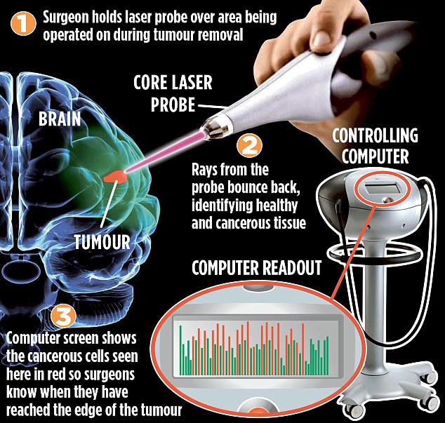
Now a British hospital is trialling a laser that bleeps like a parking sensor on a car when the scalpel gets to the edge of the cancerous areas, letting surgeons know their margins for error.In a world first, neurosurgeons at London’s Imperial College Healthcare NHS Trust have started using the pen-like probe, called the Core, which shines a near-infrared light on to a tumour and scans for subtle differences between healthy and cancerous tissue.
The device can read the differences in less than a second, and gives a warning sound if the surgeon is close to healthy tissue.
About 16,000 Britons are diagnosed with a brain tumour every year, and more children and adults under 40 die from the condition than from any other cancer. There are more than 120 types.Surgeons hope the Core, which has already proved successful in skin-cancer surgery in Canada, will allow them to remove as much of the cancerous growth as possible and also speed up operating times.The probe was used for the first time during brain surgery at Imperial last week as part of a trial that will involve 30 patients over the next year.
Senior neurosurgical trainee Babar Vaqas, who spearheaded the project to bring the Core to the UK, says: ‘There really is nothing like this at the moment.
‘Brain surgery is a very complex operation – sometimes even taking just a millimetre too much can cause real damage, such as paralysis or problems with speech. Having something that will make surgery safer and more accurate could be a real game-changer.’
One of the major obstacles at present is that the boundary between the tumour and healthy brain tissue is very hard to distinguish, even with an operating microscope.
Currently, surgeons must rely on taking several tiny cuttings of tissue to ensure they are only taking away that which is cancerous. It can take up to 90 minutes for these biopsies to be tested and for results to be fed back to surgeons, all while the patient remains in the operating theatre.
Instead of taking a physical sample, a surgeon holds the Core over the area being operated on and a beam is emitted from the tip of the probe, scanning for changes in the chemical composition of brain tissue. A tumour molecule vibrates differently from healthy molecules under laser light, causing the light to bounce back in a different way.
This is picked up by the probe, and the signal is then interpreted by a computer, giving an instant reading of whether the tissue is cancerous.
Mr Vaqas believes using the Core could shave up to 30 minutes off the average three-hour surgery time. ‘Having a device that takes less than a second to tell the difference between healthy and cancerous tissue means we could potentially avoid taking unnecessary biopsies, and also perform more surgeries in a day,’ he says.
The Core’s makers, Canadian company Verisante Technology, hopes the work at Imperial will replicate the success of a similar trial used to detect skin cancer on 1,000 patients at a hospital in Vancouver.
The Imperial trial is being part-funded by charity Brain Tumour Research Campaign, which claims research into diagnostics and treatment is ‘woefully underfunded’.
Founder Wendy Fulcher says: ‘Taking away healthy tissue can have disastrous consequences, so this probe is hugely exciting.’